🎯 Key Points
- Double circulation: pulmonary (heart↔lungs) + systemic (heart↔rest of body); SA node = pacemaker (sets the rhythm), AV node briefly delays the signal before passing it to the ventricles
- Cardiac cycle ≈0.8s: atrial systole → ventricular systole (AV valves close, "lub") → joint diastole (semilunar valves close, "dub")
- Blood pressure 120/80 mmHg (systolic/diastolic); ECG: P wave (atrial depolarisation), QRS complex (ventricular depolarisation), T wave (ventricular repolarisation)
- Blood components: RBC (haemoglobin, NO nucleus in mammals, carries O₂), WBC (immunity), platelets (clotting), plasma; ABO blood groups (IA, IB, i alleles) plus the separate Rh factor
- Frank-Starling law: greater venous return (stretching the heart more) automatically produces a stronger contraction — an intrinsic, built-in regulation that needs no nervous input at all
A single ECG cycle: the P wave marks atrial depolarisation (contraction signal), the large QRS complex marks ventricular depolarisation, and the T wave marks ventricular repolarisation (recovery) before the next cycle begins.
Composition of Blood
- Plasma: the liquid matrix (~55% of blood volume); carries proteins, nutrients, wastes, and dissolved gases
- RBCs (erythrocytes): biconcave, no nucleus in mature mammalian RBCs, packed with haemoglobin which carries O₂ (4 O₂ molecules per Hb molecule); produced via erythropoiesis, stimulated by the hormone erythropoietin (made mainly by the kidney in response to low O₂)
- WBCs (leukocytes): involved in immunity; several types (neutrophils, lymphocytes, monocytes, eosinophils, basophils)
- Platelets: cell fragments essential for blood clotting
- Blood groups: the ABO system is determined by IA, IB, and i alleles at one gene locus; the Rh factor is a separate antigen system (Rh+ vs Rh-), important in pregnancy (Rh incompatibility) and transfusions
Double Circulation and the Heart
- Pulmonary circulation: deoxygenated blood from the right ventricle → lungs (gas exchange) → oxygenated blood returns to the left atrium
- Systemic circulation: oxygenated blood from the left ventricle → entire body → deoxygenated blood returns to the right atrium
- Conduction system: SA node (pacemaker, in the right atrium, initiates each heartbeat) → spreads through atria → AV node (briefly delays the signal, allowing atria to finish contracting first) → Bundle of His → Purkinje fibres (rapidly spread the signal through both ventricles)
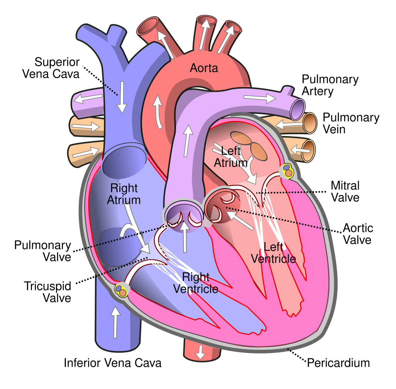
The human heart: four chambers, four valves, and the direction of blood flow. Image: Wapcaplet, CC BY-SA 3.0, via Wikimedia Commons.
Cardiac Cycle and Blood Pressure
- Cardiac cycle (about 0.8 seconds): atrial systole (atria contract, push blood into ventricles) → ventricular systole (ventricles contract, push blood into arteries; AV valves close producing the first heart sound "lub") → joint diastole (all chambers relax and fill with blood; semilunar valves close producing the second heart sound "dub")
- Cardiac output = stroke volume × heart rate; average resting cardiac output is about 5 litres per minute
- Blood pressure: systolic pressure (ventricular contraction, ~120 mmHg) over diastolic pressure (ventricular relaxation, ~80 mmHg); hypertension is sustained BP above 140/90 mmHg
- ECG: P wave (atrial depolarisation), QRS complex (ventricular depolarisation — much larger since ventricles have more muscle mass), T wave (ventricular repolarisation); Einthoven's triangle describes the standard limb-lead electrode placement used to record it
- Refractory period: cardiac muscle cells cannot be re-stimulated for a relatively long period right after contracting, which prevents tetanic (sustained) contraction and ensures the heart always relaxes fully between beats — unlike skeletal muscle, which CAN sustain tetanic contraction
- Frank-Starling law: within physiological limits, the more the heart muscle is stretched by incoming venous blood, the more forcefully it contracts — an intrinsic property of cardiac muscle requiring no external nervous signal
Blood Groups: ABO and Rh
- ABO system: based on the presence or absence of surface antigens (A and B) on RBCs and the corresponding antibodies in plasma — group A has antigen A and anti-B antibody, group B has antigen B and anti-A, group AB has both antigens and no antibodies, group O has no antigens but both antibodies
- Universal donor: group O (no antigens to be attacked); universal recipient: group AB (no antibodies to attack donor cells)
- Rh factor: a separate antigen (first found in rhesus monkeys); Rh+ individuals have the antigen, Rh- do not
- Erythroblastosis foetalis: if an Rh- mother carries an Rh+ foetus, the mother's blood may form anti-Rh antibodies (usually after the first such pregnancy); in a later Rh+ pregnancy these antibodies can cross the placenta and destroy the foetal RBCs — prevented by giving the mother anti-Rh antibodies at delivery
Coagulation (Clotting) of Blood
- A protective mechanism that seals injured vessels; a clot or coagulum is a network of insoluble fibrin threads in which blood cells are trapped
- Injured tissues and platelets release thromboplastin (a clotting factor) which, with the help of calcium ions (Ca2+), converts inactive prothrombin into the active enzyme thrombin
- Thrombin then converts soluble plasma protein fibrinogen into insoluble fibrin threads that form the meshwork of the clot
- Ca2+ ions and vitamin K (needed by the liver to make prothrombin) are essential for clotting; the whole sequence is a cascade of clotting factors
Lymph (Tissue Fluid)
- As blood passes through capillaries, some plasma leaks out into the tissue spaces as interstitial/tissue fluid; when this fluid drains into lymphatic vessels it is called lymph
- Lymph is a colourless fluid similar to plasma but with much less protein and no RBCs; it contains WBCs, chiefly lymphocytes
- Functions: returns leaked fluid and plasma proteins to the blood, carries absorbed fats from the intestine (via lacteals in the villi), and plays a key role in immunity by circulating lymphocytes and filtering pathogens through lymph nodes
Regulation of Cardiac Activity
- The heartbeat is initiated by the SA node (myogenic), but its rate and force are modulated by the nervous and endocrine systems
- The cardiac centre in the medulla oblongata controls heart activity through the autonomic nervous system
- Sympathetic nerves increase the rate of heartbeat, the strength of contraction, and cardiac output; parasympathetic (vagus) nerves decrease the heart rate, the strength of contraction, and cardiac output
- Adrenal medullary hormones (adrenaline/noradrenaline) also increase cardiac output, reinforcing the sympathetic effect during stress or exercise
Disorders of the Circulatory System
- Hypertension (high blood pressure): sustained blood pressure above 140/90 mmHg; a silent condition that strains the heart and damages arteries, kidneys, and the brain
- Coronary artery disease (atherosclerosis): narrowing of the arteries supplying the heart muscle due to deposits of calcium, fat, cholesterol, and fibrous tissue, reducing blood flow to the heart
- Angina pectoris: acute chest pain felt when the heart muscle does not receive enough oxygen, often during exertion; a warning sign of reduced coronary flow
- Heart failure: a state in which the heart is not pumping blood effectively enough to meet the body's needs (also called congestive heart failure when fluid congestion results); distinct from cardiac arrest (heart stops beating) and heart attack (sudden death of heart muscle)
🚀 NEET Advanced Edge
Why the SA node depolarises spontaneously without any external trigger: SA node cells have an unstable resting potential — a slow, automatic leak of ions (a "funny current") steadily depolarises the cell until it reaches threshold and fires an action potential on its own, repeating indefinitely; this autorhythmicity is exactly why a heart removed from the body can continue beating briefly, and why the SA node (not the brain) is the heart's true pacemaker.
Why the long cardiac refractory period is functionally essential: If cardiac muscle could be re-stimulated immediately like skeletal muscle, rapid repeated stimulation could cause sustained (tetanic) contraction — but a heart locked in continuous contraction cannot refill with blood between beats, which would be fatal; the long refractory period guarantees a full relaxation (diastole) before the next beat is even possible.
Worked reasoning: If stroke volume is 70 mL and heart rate is 72 beats/minute, calculate the cardiac output. Approach: Cardiac output = stroke volume × heart rate = 70 mL × 72 = 5040 mL/min ≈ 5.04 L/min, matching the typical resting value of about 5 L/min.