🎯 Key Points
- Resting potential −70mV (Na⁺/K⁺ pump: 3 Na⁺ out, 2 K⁺ in); Action potential: Na⁺ in (depolarisation to +40mV) → K⁺ out (repolarisation); threshold ≈ −55mV; follows all-or-nothing law
- Myelin (Schwann cells in PNS, oligodendrocytes in CNS) enables FAST saltatory conduction by jumping between Nodes of Ranvier
- Cerebrum=thinking/voluntary movement; Cerebellum=balance/coordination; Medulla=involuntary (breathing, heart rate); Hypothalamus=homeostasis+controls pituitary
- Reflex arc: Receptor→Sensory neuron→Spinal cord (interneuron)→Motor neuron→Effector — does NOT require the brain, which is why reflexes are faster than voluntary responses
- Sympathetic = fight-or-flight (↑heart rate, ↑pupil dilation); Parasympathetic = rest-and-digest (↓heart rate, ↑digestion) — antagonistic on the same organs
Neuron Structure
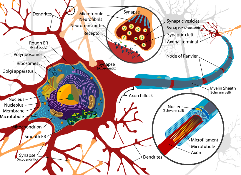
Structure of a neuron. Image: LadyofHats (Mariana Ruiz), Public Domain, via Wikimedia Commons.
- Dendrites → Cell body (soma) → Axon → Axon terminals
- Myelin sheath: Schwann cells (PNS), oligodendrocytes (CNS); speeds conduction (saltatory)
- Nodes of Ranvier: gaps in myelin sheath
Nerve Impulse
- Resting potential: -70 mV (Na+/K+ pump: 3 Na+ out, 2 K+ in)
- Action potential: Na+ rushes in (depolarization to +40 mV) → K+ rushes out (repolarization)
- All-or-nothing law; threshold: -55 mV
- Synapse: neurotransmitters (acetylcholine, noradrenaline, dopamine, serotonin) cross synaptic cleft
Brain Regions
- Cerebrum: thinking, memory, speech, voluntary movement; largest region
- Cerebellum: balance, posture, fine motor coordination
- Medulla oblongata: breathing, heart rate, vomiting, swallowing
- Hypothalamus: thermoregulation, hunger, thirst, sleep, controls pituitary
- Thalamus: sensory relay station
- Limbic system: emotions, memory (hippocampus)
Sense Organs
- Eye: cornea → lens → retina (rods-dim light, cones-colour); fovea (sharpest vision); optic nerve
- Ear: outer (pinna) → tympanum → ossicles (malleus, incus, stapes) → cochlea (hearing); semicircular canals (balance)
Reflex Arc
Receptor → Sensory neuron → Spinal cord (interneuron) → Motor neuron → Effector (muscle/gland)
PNS: Somatic (voluntary) + Autonomic (sympathetic vs parasympathetic)
Types of Neurons
- Sensory (afferent) neurons: carry impulses from receptors toward the CNS
- Motor (efferent) neurons: carry impulses from the CNS to effectors (muscles/glands)
- Interneurons (association/relay neurons): connect sensory and motor neurons, mostly within the CNS
- Structurally: multipolar (many dendrites, one axon; most common, e.g. motor neurons), bipolar (one dendrite, one axon; retina), unipolar (single process; sensory ganglia)
Synaptic Transmission
- Synapse: junction between the axon terminal of one neuron and the dendrite of the next; electrical synapse (direct, fast, via gap junctions) vs chemical synapse (via neurotransmitters, most common)
- At a chemical synapse: action potential reaches axon terminal, triggers Ca2+ influx, synaptic vesicles fuse with the membrane and release neurotransmitter into the synaptic cleft, which binds receptors on the postsynaptic membrane and generates a new impulse
- Neurotransmitters are then either broken down by enzymes (e.g. acetylcholinesterase degrades acetylcholine) or taken back up (reuptake), ending the signal
- Synapses can be excitatory (depolarise postsynaptic membrane) or inhibitory (hyperpolarise it)
Organisation of the Nervous System
- Central Nervous System (CNS): brain and spinal cord; site of information processing and control
- Peripheral Nervous System (PNS): all nerves connecting CNS to the rest of the body; divided into cranial nerves (12 pairs, from brain) and spinal nerves (31 pairs, from spinal cord)
- PNS is functionally divided into the somatic nervous system (voluntary control of skeletal muscle) and the autonomic nervous system (involuntary control of internal organs)
- Sympathetic division: prepares body for stress ("fight or flight"); increases heart rate, dilates pupils; Parasympathetic division: promotes rest and digestion ("rest and digest"); slows heart rate, stimulates digestion; the two usually act antagonistically on the same organ
The Human Brain: Forebrain, Midbrain and Hindbrain
- Forebrain: cerebrum (largest part; two cerebral hemispheres joined by the corpus callosum; controls voluntary movement, intelligence, memory and speech), thalamus (major sensory and motor relay centre) and hypothalamus (controls body temperature, hunger, thirst, sleep and the pituitary gland)
- Midbrain: located between the thalamus/hypothalamus and the pons; its dorsal part bears four rounded lobes, the corpora quadrigemina, which control visual and auditory reflexes
- Hindbrain: pons (connects brain regions, helps regulate breathing), cerebellum (coordinates precise voluntary movements, posture and balance) and medulla oblongata (controls respiration, cardiovascular reflexes and gastric secretions)
- The midbrain and hindbrain together form the brainstem, the connection between the brain and the spinal cord
- The cerebral cortex is folded to increase surface area and contains motor, sensory and association areas; the inner white matter carries myelinated nerve fibres
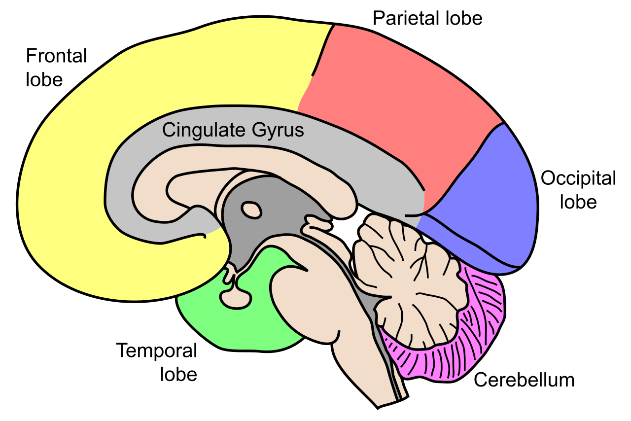
Mid-sagittal view of the human brain showing the four cerebral lobes and the cerebellum. Image: NEUROtiker, CC BY-SA 3.0, via Wikimedia Commons.
The Eye and Mechanism of Vision
- The eyeball wall has three layers: outer sclera (transparent cornea in front), middle vascular choroid (thickened in front to form the ciliary body and iris) and inner retina
- The iris controls the size of the pupil and hence how much light enters; the lens, held by ligaments on the ciliary body, focuses light on the retina (accommodation)
- The retina bears photoreceptors: rods (contain the pigment rhodopsin/visual purple; function in dim light and give black-and-white twilight vision) and cones (three types sensitive to red, green and blue; give colour vision and bright-light vision)
- The fovea (macula lutea) is the point of sharpest vision, densely packed with cones; the blind spot is where the optic nerve leaves the eye and has no photoreceptors
- Mechanism: light focused on the retina causes breakdown of photopigments, generating impulses in the photoreceptors; these pass through bipolar and ganglion cells to the optic nerve and reach the visual cortex, where the image is interpreted
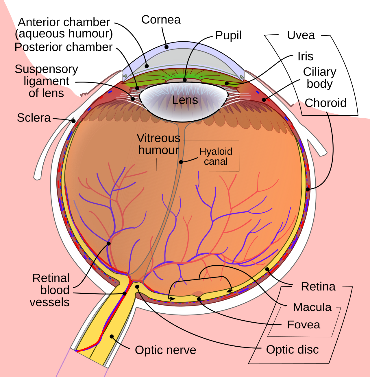
Cross-section of the human eye: light passes through the cornea and lens to focus on the retina, where rods and cones lie and the fovea gives sharpest vision. Image: Rhcastilhos and Jmarchn, CC BY-SA 3.0, via Wikimedia Commons.
The Ear and Mechanism of Hearing
- Outer ear: the pinna collects sound waves and directs them through the external auditory canal to the tympanic membrane (eardrum)
- Middle ear: three ear ossicles — malleus, incus and stapes — amplify and transmit vibrations; the Eustachian tube connects the middle ear to the pharynx and equalises air pressure on both sides of the eardrum
- Inner ear: the fluid-filled coiled cochlea houses the organ of Corti with sensory hair cells (hearing); the vestibular apparatus (three semicircular canals plus utricle and saccule) maintains balance and equilibrium
- Mechanism: vibrations pass eardrum → ossicles → oval window → cochlear fluid, setting up ripples in the basilar membrane; hair cells of the organ of Corti bend against the tectorial membrane, generating impulses carried by the auditory nerve to the auditory cortex, where sound is recognised
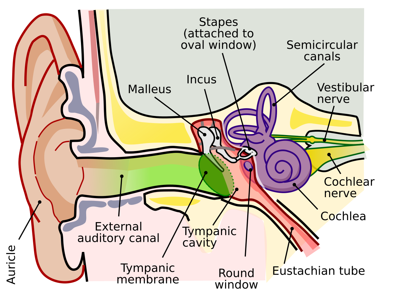
The human ear: the outer ear funnels sound to the eardrum, the middle-ear ossicles (malleus, incus, stapes) amplify it, and the cochlea of the inner ear converts the vibrations into nerve impulses. Image: Lars Chittka and Axel Brockmann, CC BY 2.5, via Wikimedia Commons.
Reflex Action and Types of Reflexes
- A reflex action is a rapid, automatic, involuntary response to a stimulus, mediated by the spinal cord without conscious involvement of the brain
- The pathway is the reflex arc: receptor → afferent (sensory) neuron → CNS (spinal cord, usually via an interneuron) → efferent (motor) neuron → effector (muscle or gland)
- Monosynaptic reflex: e.g. the knee-jerk (stretch) reflex, with a single synapse between the sensory and motor neurons; polysynaptic reflex: e.g. the withdrawal reflex on touching a hot object, involving one or more interneurons
- Reflexes are protective and are faster than voluntary responses because the impulse does not need to travel up to the brain before a response is produced
🚀 NEET Advanced Edge
Why saltatory conduction is faster: In myelinated axons, the action potential "jumps" from one Node of Ranvier to the next (ion channels are concentrated only at the nodes), rather than regenerating continuously along the entire membrane as in unmyelinated fibres — this is why demyelinating diseases (e.g. multiple sclerosis) drastically slow nerve conduction.
Refractory period and impulse direction: Immediately after an action potential fires, the Na⁺ channels at that point briefly cannot reopen (absolute refractory period) — this ensures the impulse travels in ONE direction only (away from the already-fired region), preventing back-propagation along the axon.
Drugs/toxins acting at the synapse: Acetylcholinesterase inhibitors (e.g. some nerve agents, certain pesticides) prevent breakdown of acetylcholine, causing continuous stimulation of the postsynaptic membrane — connecting basic synapse mechanism to toxicology, a common applied NEET question style.